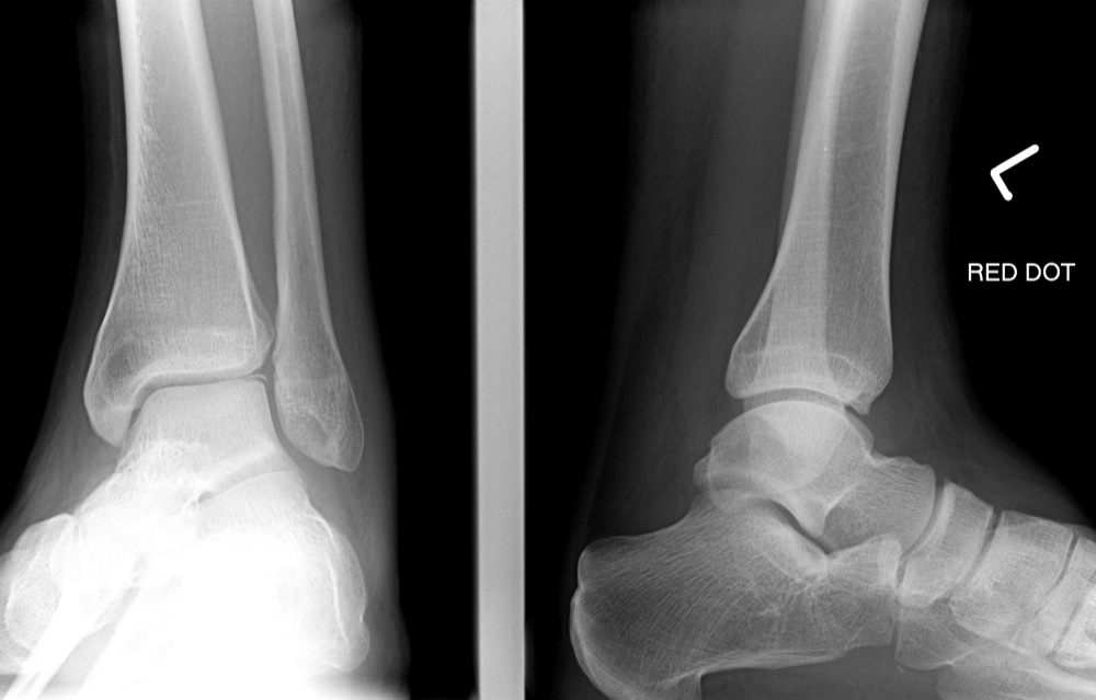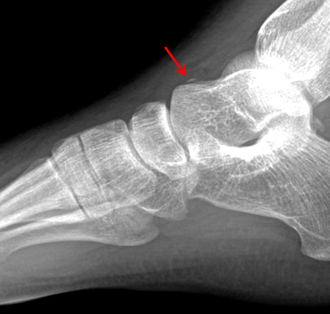

Despite these advances, functional outcomes remain poor in a subset of severely injured patients, making further research imperative. The suggested mechanism is thought to be a consequence of forced dorsiflexion and inversion of a fixed pronated foot. In general, talar neck and body fractures should be treated operatively if the fracture is displaced more than 1 to 2 mm. An unstable Talus Fracture occurs when a piece of bone is displaced from its attachment. Lateral process fractures are usually a result of high-energy injuries. There are five major types of talus fractures: talar head, neck, body, process or tubercle, and osteochondral fractures, and each type has site-specific indications for operative treatment.

A variety of new treatments for these complications have been proposed, including vascularized autograft, talar replacement, total ankle arthroplasty, and improved salvage techniques, permitting some patients to return to a higher level of function than was previously possible. The most common type of fracture to the Talus is to the neck, which attaches the body to the neck of the talus bone, and this can be a stable or unstable fracture. Despite this modern technique, osteonecrosis and osteoarthritis remain common complications. The talus, a small bone in the hindfoot: Articulates with the calcaneus and the remainder of the midfoot bones. Percutaneous screw fixation has also demonstrated success in certain patterns. The widely preferred approach for many talar neck and body fractures is a dual anterior incision technique to achieve an anatomic reduction, with the addition of a medial malleolar osteotomy as needed to visualize the posterior talar body. Fractures of the head, neck, or body regions have the potential to compromise nearby joints and impair vascular inflow, necessitating surgical treatment with stable internal fixation in many cases. Treatment of arthritis includes activity modifications, ankle braces, and either ankle joint fusion or replacement.The talus is unique in having a tenuous vascular supply and 57% of its surface covered by articular cartilage. If the cartilage is damaged, the bones will be forced to rub against each other without that protection. Since most of the talus is covered with cartilage, bones are allowed to move smoothly against each other. The anterolateral approach is usually employed in combination with the anteromedial approach to gain access to any displaced talar neck fracture. Jorge Bowakim, Javier Lpez-Goenaga, Javier Mayo, Julin del Ro. This will reveal if the talus bone has a good blood supply.Įven if the bones heal well, arthritis may still develop. Osteochondral Autologous Graft in the Treatment of an Open Talar Fracture: A Case Report. During healing, the physician may request X-rays or an MRI to be done. Immediate first aid for a talar fracture is to apply a well-padded splint around the back of the foot and leg from the toe to the upper calf. These may include not being able to use your foot normally, arthritis, chronic pain or collapse of the bone. The patient will not be allowed to put any weight on the foot for at least two to three months. A talar fracture that doesn't heal properly will create problems for you later. Any small fragments of bone discovered during this procedure will be removed and bone grafts will be used to help restore the shape of the joint.Īfter surgery, the patient will then be placed in a cast for approximately eight to twelve weeks. The orthopaedic surgeon realigns the broken bone with metal screws placed inside the bone. M ost fractures of the talus do require surgery to reset the bone and help minimize later complications.


 0 kommentar(er)
